The major health effect of uranium exposure has been reported to be chemical kidney toxicity, functional and histological damages being mainly observed in proximal tubule cells. Uranium enters the proximal tubule as uranyl-bicarbonate or uranyl-citrate complexes. The aim of our research is to investigate the mechanisms of uranium toxicity, intracellular accumulation and repartition after acute intoxication of rat renal proximal tubule epithelial cells, as a function of its chemical form.
Microscopic observations of renal epithelial cells after acute exposure to uranyl-bicarbonate showing the presence of intracellular precipitates as thin needles of uranyl-phosphate localized in cell lysosomes have been published. However the initial site of precipitates formation has not been identified yet: they could either be formed outside the cells before internalization, or directly inside the cells.
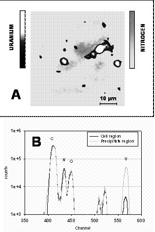
Uranium solubility as a function and initial concentration was specified by ICP-MS analysis of culture media. In parallel, uranium uptake and distribution in cell monolayers exposed to U-bicarbonate was investigated by Nuclear Microprobe analyses.
The present study shows that a non-negligible proportion of uranyl-bicarbonate solutions precipitates when diluted in classical cell culture medium. Various experimental techniques have been used, and nuclear microprobe analyses permitted to establish a correlation between uranium incorporation and toxicity. Moreover, nuclear microprobe imaging analyses revealed that uranium may be present in both precipitate and soluble forms inside the cells. An optimized RBS arrangement appeared to be more efficient than standard PIXE setups when measuring heavy metal thin layers in an organic matrix. The presence of these two U chemical forms inside the cells can either come from initial soluble U internalization and then partial precipitation as described elsewhere, or more probably from two parallel internalization paths. U internalization mechanisms will be further studied in order to define the most toxic uranium chemical form, i.e. the soluble or precipitated form.