|
|
Neutron radiography : non destructive testing
For many years, most of the Material Testing Reactors of French Atomic Commission (CEA) have been equipped with Neutron Radiography facilities.
Industrial NR activities are carried out by a small team meeting customers requirements in term of costs and schedule.
Principle
Neutron Radiography is an imaging technique which provides images similar to X-ray radiography. The difference between neutron and X-ray interaction mechanisms produce significantly different and often complementary information. While X-ray attenuation is directly dependent on atomic number, neutrons are efficiently attenuated by only a few specific elements. For example, organic materials or water are clearly visible in neutron radiographs because of their high hydrogen content, while many structural materials such as aluminium or steel are nearly transparent. The next table shows how most materials behave when placed in the path of a neutron beam.
Industrial applications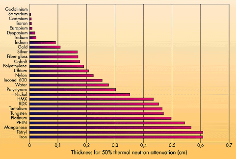
|
|
At the present time, Neutron Radiography is one of the main NDT technique able to satisfy the quality-control requirements of explosive devices used in space programmes. Most of the detonating devices of the Ariane space programme have been systematically submitted to NR examination at the CEA facilities for more than 20 years. The detection of cracks of 0.1 mm thickness in the explosive charge is common and the efficiency of the technique enables easily distinguishes differences in the compression of the explosive even through different metallic containers such as lead, aluminium or steel. The ability to detect compounds containing hydrogen atoms is also used to inspect oil levels and insulating organic materials. Neutron radiography also facilitates the checking of adhesive layers in composite materials ,surface layers (polymers, varnishes etc). All types of O-rings and joints containing hydrogen can be observed even through a few centimetres thickness of steel. |
|
|
|
|
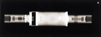
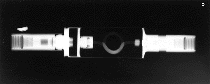
Comparison of neutron radiography and X-ray radiography of an ARIANE cartridge fuse (DASSAULT-AVIATION) |
Facilities
|
|
Two nuclear research reactors in Saclay carry out industrial NR inspections. The main facility is located at the neutron guide end of the ORPHEE reactor. This reactor is currently in operation 250 days a year. The beam quality has a very high intensity free from parasitic gamma rays. The beam has a cold neutron component which produce high contrast radiographs. The second facility is installed at the ISIS reactor, it is used when the ORPHEE reactor is shut down. |
Conditions of access
Equipment
French references
Neutron radioscopy : dynamic neutron imaging
|
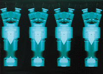
Neutron radioscopy examination of the flow of oil (CEA DRN /DRE)
|
Neutron radioscopy consists of the continuous visualisation of the attenuation of a neutron beam using a " real time " detector. Whereas Neutron Radiography is imaged on a photographic film, radioscopy uses a scintillator and a video camera.
|
Principle
|
Colour simulation of the flow of a refrigerant (TOTAL/CEA)
|
Neutron Radiography is an imaging technique which provides images similar to X-ray radiography. Neutron interactions with matter can be divided into scattering and absorption. Neutrons are able to detect elements containing hydrogen atoms through metallic containers. The information provided by spatial and temporal beam attenuation is recorded on magnetic media via analogic or digital signals.
|
Applications
Most of the applications known so far, consist in visualisation of fluids moving through metallic containers. In fact, hydrogen atoms included in such fluids are detected by neutrons. The main fields where dynamic neutron imaging has been used are:
* oil lubrication (engines, gear boxes...)
* fuel behaviours (carburettors, injectors...)
* two-phase flow (heat exchangers, condensers, steam generator tubes...)
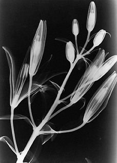
* transfer and migration of fluids into porous media (wetting of soils, pollution migration, plants growing...)
|
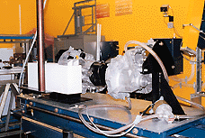
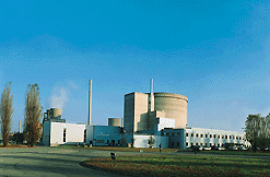
Orphée reactor (CEA DRN/DRE) |
Two neutron beams extracted from Saclay research reactors can be used for neutronoscopy. The most practicable is located at the end of a neutron guide of the Orphee reactor. The beam cross section is limited to 50 x 30 mm 2 but the flux level and the spectrum (free from gamma contribution) allow images of great quality to be obtained. The second facility has a 18 x 24 cm 2 neutron beam on the ISIS reactor. |
|
|
Access conditions Equipment
|
French references